
Five labs with one goal: saving sight
Miami researchers are working to understand, prevent, and repair vision loss
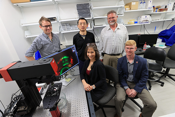
Five labs with one goal: saving sight
Miami researchers are working to understand, prevent, and repair vision loss
Vision loss and blindness caused by cataracts, glaucoma, diabetic retinopathy, and macular degeneration affect approximately one billion people globally, according to the World Health Organization. These conditions occur when the lens or retina of the eye has been damaged by aging, genetics, medical conditions, or injuries.
At Miami University, researchers are working to understand how eyes develop and how disease takes hold, how damage might be repaired, and how problems can be caught sooner. Their work spans biology and engineering, from genes and model organisms to imaging technology.
Mike Robinson: How one gene shapes the eye
development and retina regeneration. Currently, his lab is focusing on FOXE3, a transcription factor gene that is key for lens development.
Transcription factors are proteins that control the expression of other genes. By switching genes on or off, they direct how cells grow and function in particular parts of the body. When FOXE3 is mutated, humans can develop microphthalmia, a birth defect in which one or both eyes do not fully develop and are abnormally small, resulting in vision impairment or blindness, depending on its severity.
Robinson’s lab wants to gain insight into which genes FOXE3 controls to make a lens and how it does so. To pinpoint FOXE3’s targets, the lab uses transcriptomics, a technique that reveals which genes are active under specific conditions, such as developmental stages, tissue types, and disease states. The early results point to a surprising role for the gene.
“We think one of the major roles of FOXE3 is not to turn genes on to make cells into lenses, but to turn genes off that would otherwise make neural tissue, particularly retina,” Robinson said. Because the lens and retina develop from neighboring tissue, cells need switches that lock in one identity and keep the other off. Understanding how that decision is made could shed light on why lens development fails when FOXE3 is mutated, and could inform how other tissues develop.
Robinson and colleague Chung Liang, professor of Biology, recently received $467,969 from the National Eye Institute for their project "FOXE3 in Creating Lens Chromatin Architecture and Transcriptional Identity."
Mark Charlton-Perkins: A window into tumor growth
Neurofibromatosis type 1 (NF1) is a common inherited genetic disorder that can cause tumors to develop throughout the nervous system. Approximately 15 to 20% of children with NF1 develop optic pathway gliomas, tumors that affect the visual pathway connecting the eyes to the brain. While many of these tumors remain stable, others can lead to significant vision loss. Why some children develop these tumors while others do not remains an important unanswered question.
Mark Charlton-Perkins, assistant professor of Biology, is investigating the genetic factors that contribute to more severe forms of NF1. His research is part of a multi-year National Institute of Health-funded collaboration between Miami University, Cincinnati Children’s Hospital Medical Center, and Lurie Children’s Hospital of Chicago.
By combining genetic information with patients identified through these clinical partnerships with zebrafish models of NF1, Charlton-Perkins and his collaborators are working to understand why some individuals develop more severe disease. His laboratory uses genetic engineering to investigate how changes in gene activity contribute to tumor formation and progression.
Ultimately, the researchers hope to identify genetic factors that could help predict which patients are at greater risk of developing optic pathway gliomas and uncover new therapeutic targets that could lead to more personalized treatments.
Charlton-Perkins’s laboratory has been awarded more than $1 million in external research funding to support this and related research programs.
Katia Del Rio-Tsonis: Newt secrets for eye tissue regeneration
Newts have long fascinated researchers with their ability to regenerate limbs and organs after damage, though the cellular mechanisms behind these processes aren’t well understood. Katia Del Rio-Tsonis, professor of Biology, wants to crack the code for regenerating the lens and retina, hoping to help pave the way for therapies that restore vision lost to blinding conditions.
Using animal models like Spanish newts and embryonic chicks, Del Rio-Tsonis’s lab can test the animals’ regenerative abilities by removing the lens or the retina and manipulating genes and signaling pathways (that have been identified by transcriptomic analysis) as they regenerate. Through these studies, the lab aims to identify key genes underlying regenerative capacity that could eventually be targeted to promote regeneration in higher vertebrates, including humans.
Del Rio-Tsonis believes that macrophages — white blood cells of the immune system that respond to injury signals, destroy pathogens, and help with wound healing — play a role in whether regeneration happens.
“Our hypothesis is that you need injury signals, but they have to be acute, not chronic. Persistent inflammation can impair tissue repair and contribute to scarring in many diseases. Newts can regulate and resolve the inflammatory response after injury in ways that support regeneration with minimal scarring,” Del Rio-Tsonis said. She thinks key inflammatory signals could be used to reprogram damaged cells in the eye to grow new cells or neurons.
Del Rio-Tsonis, who was named a 2026 University Faculty Scholar in recognition of sustained excellence in research, has received more than $9.7 million in research funding since joining Miami in 1999.
Andrew Jones: Bacteria as tiny compound factories
What does metabolic engineering have to do with eye research? For Andrew Jones, associate professor of Chemical, Paper, and Biomedical Engineering, the answer is a supply chain: His lab makes compounds that other Miami labs use in their studies.
“The idea is that nature provides a diversity of drug candidates, but many of the native organisms can not be cultivated to produce the drugs at a suitable scale and purity,” Jones said. “We’re taking metabolic pathways from mushrooms, toads, and plants that make these natural psychedelics and we’re genetically encoding these pathways in E. coli to give it the ability to produce these compounds in a scalable and reproducible way.”
The bacteria are grown under controlled and optimized conditions to maximize drug biosynthesis capabilities and then the drug is purified and crystallized for use in other labs. The compounds produced range from classical psychedelics, like psilocybin, to non-hallucinogenic derivatives which are mostly unstudied in the scientific literature.
Inflammation contributes to many diseases, including eye diseases. Some psychedelics have anti-inflammatory properties, and because chronic inflammation can block healing, these compounds could help support tissue regeneration and make treatments for eye diseases more effective. That idea echoes Del Rio-Tsonis’s hypothesis that regeneration depends on resolving inflammation.
Jones said he is interested in making the compounds more reliable, sustainable, cheaper, and reproducible. His lab uses a range of analytical chemistry equipment to help determine whether their genetic modifications improved or worsened the end product. If a lab requests a compound for a specific application, Jones’ team can adjust their synthesis and purification processes accordingly.
Hui Wang: Sharper images for earlier diagnosis
Imagine getting an eye screening at the pharmacy, with no on-site technician needed. Hui Wang, associate professor of Chemical, Paper, and Biomedical Engineering, wants to make that possible by improving the imaging technology used to diagnose eye diseases.
His BioPhotonics lab concentrates on optical coherence tomography (OCT), a noninvasive imaging technique that uses light waves to take images of the retina and optic nerve. Physicians can use retina images to diagnose and monitor ocular diseases such as glaucoma and age-related macular degeneration. Beyond ophthalmology, the emerging field of oculomics explores how ocular imaging can reveal biomarkers of systemic diseases, including cardiovascular, metabolic, and neurodegenerative disorders.
However, a major challenge in eye care is that vision loss caused by many retinal diseases is irreversible, Wang said. Early detection is therefore critical, as it allows physicians to intervene before significant damage occurs and potentially slow disease progression. Achieving this requires high-resolution retinal imaging capable of detecting subtle changes at the earliest stages of disease.
This is where adaptive optics OCT comes in. Images taken with this technology capture the retina and ocular tissue at the cellular level and correct distortions in the eye, allowing retinal diseases to be diagnosed earlier. Wang’s lab aims to develop the next generation of adaptive OCT technology by integrating computational optical imaging, bringing high-resolution retinal imaging closer to routine clinical practice.
In another research direction, Wang’s lab aims to make retinal imaging more accessible to the general population. Many retinal diseases require frequent follow-up examinations, but providing regular monitoring to large populations remains a major challenge. To address this, Wang’s team is developing autonomous tele-OCT systems that could be deployed in community centers and pharmacies, allowing patients to undergo retinal imaging without human assistance. As these systems will generate large volumes of imaging data, AI will play an essential role in detecting abnormalities, assisting diagnosis, and identifying patients who need referral to eye specialists. Wang believes such community-based retinal imaging could become a reality within the next five to 10 years. Wang’s lab has been funded over 1.5 million from DoD and NIH and Wang received the 2025 University Faculty Scholar Award.
For the one billion people living with vision loss, the answers won’t come from a single lab. These projects by Miami researchers differ in method, but together they span the full arc of eye disease: how it begins, how it might be reversed, and how it can be caught early. Together, they offer steps toward a future in which vision loss no longer has to be permanent.