
Small molecule, big consequences: A look inside Miami’s group of protein researchers
Four Miami researchers are unraveling how a single molecule can shape human health outcomes
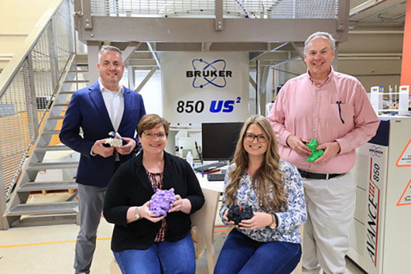
Small molecule, big consequences: A look inside Miami’s group of protein researchers
Four Miami researchers are unraveling how a single molecule can shape human health outcomes
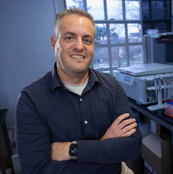
Rick Page: Protein quality control and antibiotic resistance
Rick Page, interim vice president for Research and Innovation and professor of Chemistry and Biochemistry, focuses on protein quality control and antibiotic resistance within his lab.
When proteins become misfolded, a specialized molecule called a chaperone will help refold them or guide them to be recycled back into their original amino acids to make proteins elsewhere. Cancer cells, Page said, have gotten very good at hijacking this system — growing quickly by putting their chaperones to work.
That system is the focus of the protein quality control arm of his lab, which studies how proteins in cancer cells work to lay the foundation for future therapeutics. Using synthetic proteins, the lab creates mutations that control how well-folded a protein is, then observes how chaperones respond by either refolding or recycling it.
“A strategy for anti-cancer therapeutics is to disrupt that process in cancer cells, and the idea is if you do that, it can help kill or weaken them. It can sensitize them to lower concentrations of typical anti-cancer drugs,” Page said.
The antibiotic resistance arm of the lab looks at proteins that perform a hydrolysis reaction on antibiotics like ampicillin, amoxicillin, and penicillin, making them inactive and unable to kill bacteria. They study the structure of the bacteria’s proteins and their dynamics (movement and changes) to better understand how they work and develop inhibitors for them.
“It’s a way bacteria have evolved to have resistance. It’s a huge problem in the healthcare industry across the world,” Page said.
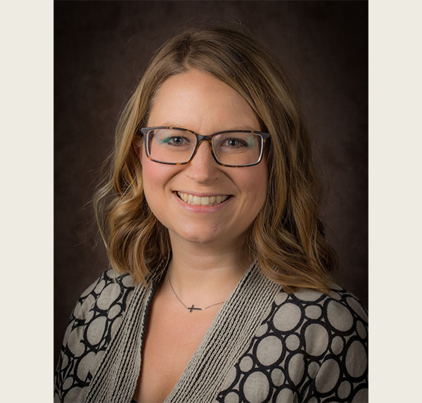
Andrea Kravats: Chaperones and misfolded proteins
Misfolded proteins tend to clump together, known as aggregation, and those aggregates cause neurodegenerative diseases, cancer, and glaucoma. Andrea Kravats, associate professor of Chemistry and Biochemistry, studies chaperones located in the endoplasmic reticulum of cells that work together to prevent or inadvertently help these diseases.
“The problem is that chaperones aren’t always able to discriminate between good and bad proteins,” Kravats said. “If you have a disease-causing protein that is mutated and is never going to attain its shape, the chaperones will interact with it, stabilize it, and help carry out those bad effects.”
Pinning down what features chaperones look for in misfolded proteins, and what they need to function, could point the way toward new therapeutics for these diseases.
Her computational lab runs simulations — some of which have been running continuously for two years — on the 3D structures of proteins, tracking the environmental factors that trigger the shape changes at the root of aggregation-linked disease.
Her experimental lab tests how chaperones work together to remodel misfolded proteins, mapping each step of the remodeling pathway so it can be targeted with drugs. Right now, the lab is using firefly luciferase — the protein that lets the bug’s abdomen glow — as a stand-in for this process. The protein is denatured so it misfolds and stops lighting up, then chaperones are added and tracked as they refold it and restore its luminescence, giving researchers a visible readout of how well a given chaperone intervention works.
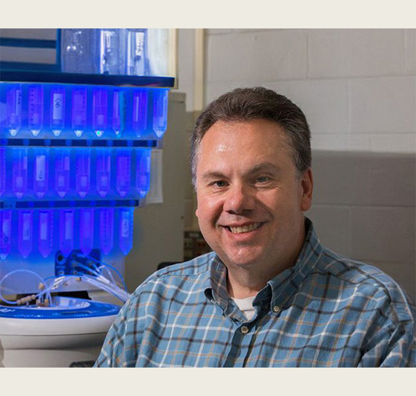
Gary Lorigan: Proteins and cardiac potassium channels
Eight different proteins — some of which bind to the potassium channels in our hearts — are the focus of University Distinguished Professor of Chemistry and Biochemistry Gary Lorigan’s lab, where he studies their structure and function.
One such mutated protein is connected to Long QT Syndrome, a heart signaling disorder that delays the electrical recharging of the heart between beats, which can cause fainting without warning, seizures, heart palpitations, cardiac arrest, or even sudden death.
His lab collaborates with colleagues at Vanderbilt University, creating protein mutations which are then sent to a Vanderbilt lab and injected into frog oocytes. By tracking how well the potassium channels are functioning within the oocytes, researchers could create drugs that can help manage Long QT Syndrome and other heart diseases.
Carole Dabney-Smith: Protein movement across membranes in plant cells and bacteria
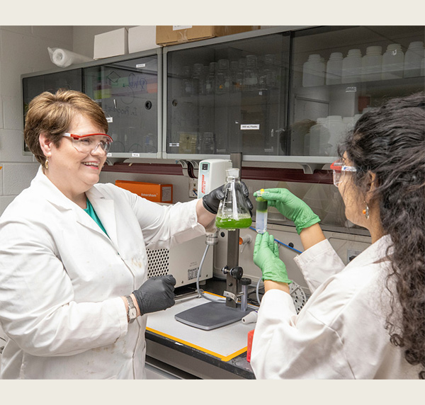
Professor of Chemistry and Biochemistry Carole Dabney-Smith’s lab is markedly different from the rest of the group as her lab studies how proteins move from one space to another in a plant cell’s chloroplast and how the proteins that make up the membrane “doorways” work to let other proteins through.
“If a protein is made in the kitchen and needs to get to the den, they have to go through a door, and that process is called translocation,” Dabney-Smith said, comparing the spaces within a cell to rooms within a house.
Dabney-Smith wants to pin down the mechanism for how and when the door opens. This is tested by isolating the chloroplast from the rest of the cell, giving it a protein that needs to cross a membrane, and labeling that protein so they can follow as it moves. There are all kinds of chemical signals that indicate when the “door” needs to open and close, and what shape the proteins need to be to successfully navigate through the doorway.
This doorway research has applications for both agriculture and antibiotics because the same doorways are found in plant chloroplasts and bacterial membranes. Engineering plants to control how proteins move through membranes could make them hardier, helping them survive — and yield better — in tougher growing conditions. The same knowledge could also lead to the development of drugs that stop the doorway mechanism from working in bacteria, making pathogenic bacteria less effective at infecting humans and livestock while making existing antibiotics work better when taken together.
Though each lab is chasing a different end goal, their starting point is the same. Together, they’re building a shared portrait of how one of the body’s most fundamental building blocks works — and how a single molecule going wrong can ripple outward into nearly every corner of human health and wellness.